
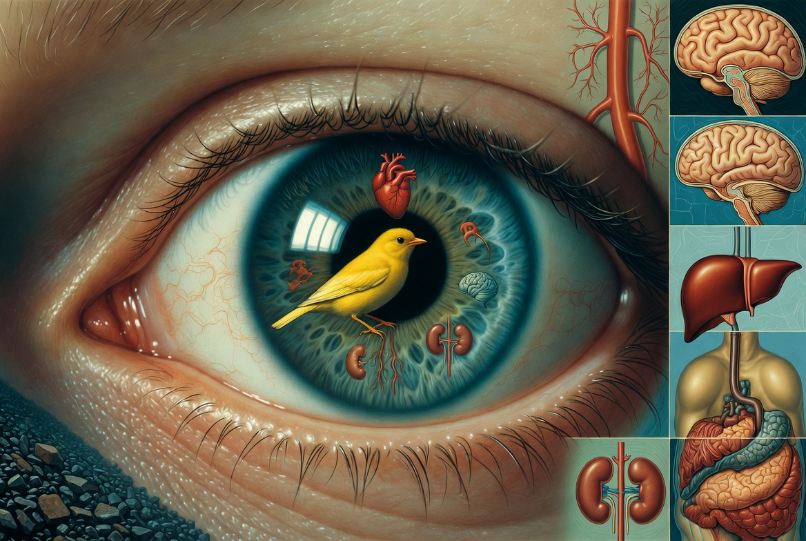
Eye Test for Systemic Problems
Take the form to your eye doctor (link below).
$10.00
FrugalDoc recommends getting a comprehensive eye exam every 5 years after age 40. There are 3 key tests to obtain.
OCT: The OCT instrument accurately and precisely measures the health of your retina. This is important because the retina IS part of your brain. Importantly, this instrument is capable of detecting the earliest retinal changes that are often reflected in the brain. In this regard, it is superior to a brain MRI.
Optical Coherence Tomography (OCT) provides a convenient “window to the brain” by directly imaging retinal neurodegeneration and neurological changes. It excels at detecting structural loss of neurons and axons but works best as part of a broader assessment. It is a noninvasive imaging technique that uses near-infrared light to produce high-resolution, cross-sectional (3D) images of the retina. It measures the thickness and structure of retinal layers with micrometer-level precision.
The specific tissue it measures is the retinal nerve fiber layer (RNFL).
The Key Way OCT Detects Neurodegeneration
Retinal Nerve Fiber Layer (RNFL) Thinning
Measures the thickness of axons from retinal ganglion cells.
Thinning indicates axonal loss, a hallmark of neurodegeneration.
Commonly seen in Alzheimer’s disease (AD), Parkinson’s disease (PD), multiple sclerosis (MS), and other neurological conditions like Major Depressive Disorder, Bipolar Disorder, and Persistent Depressive Disorder.
OCT Angiography (OCTA) is the highest level OCT that fewer eye doctors have. This instrument measures everything the standard OCT does, and also gives a detailed look at the health of your vessels. Thus, OCTA can...
Assesses retinal microvasculature (vessel density and perfusion).
Reduced vessel density or enlarged foveal avascular zone can indicate vascular contributions to neurodegeneration (common in AD and PD).
It is also an indicator of whole-body small vessel (capillary) health and thus is arguably the earliest way to "see" vascular and heart disease.
Fundus Camera: A fundus camera is a specialized low-power microscope with a camera attachment that captures high-resolution color photographs of the fundus — the interior surface of the eye, including the retina, macula, optic disc, and retinal blood vessels.
IMPORTANT: The vessels it measures are in a capillary network called the Chorid. This is the most dense set of vessels anywhere in the body - by far, because it is supporting the most active tissue in your body, the RNFL - which is your "photographic plate."
Detecting Retinal Vessel Disease (and Systemic Vascular Issues)
The retina is the only place in the body where small blood vessels (microvasculature) can be directly visualized non-invasively. Fundus images reveal changes that reflect both local and systemic vascular health.
Common findings:
Arteriolar narrowing, silver/copper wiring (thickened vessel walls).
Arteriovenous nicking (veins compressed where crossed by arteries).
Hemorrhages, microaneurysms, cotton-wool spots (nerve fiber layer infarcts), hard exudates.
Vessel tortuosity, dilation, or reduced density.
These indicate hypertensive retinopathy, diabetic retinopathy, or generalized atherosclerosis and microvascular damage.
Linking to Heart Disease and Cardiovascular Risk
Retinal microvascular changes often mirror similar damage in the brain, heart, and kidneys. Fundus photography (and especially AI-enhanced analysis) helps assess cardiovascular risk.
Slit Lamp Microscope. This inexpensive instrument scans the lens of your eye for opaque structures that are NOT present in a healthy eye and body.
IMPORTANT: The slit lamp microscope can detect and measure two (2) types of cataracts. The common cataract is called a "nuclear" cataract, and a "cortical" cataract that appears on the edge (cortex) of the lens (does not impact vision). They are both predictors of future chronic degenerative disease - but each provides different insights into your future health.
NUCLEAR CATARACT
The key study on people with and without nuclear cataract, the common cataract that impacts your central vision, is the Age-Related Eye Disease Study (AREDS) study conducted by the NIH. AREDS was a large, multicenter, prospective study (and randomized trial) involving about 4,757 participants aged 55–80 years.
Key Findings on Cataract and Heart Disease
The study found that common age-related eye conditions, including cataract (specifically nuclear opacity and history of cataract surgery), were linked to higher overall mortality and, in some cases, cardiovascular mortality.
Specific associations included:
Nuclear opacity (a common type of age-related cataract): Associated with a 40% increased risk of all-cause mortality.
Cataract surgery (indicating significant cataract - not the surgery component): Associated with a 55% increased risk of all-cause mortality and notably linked to cancer deaths in the analysis.
Advanced macular degeneration: Strongly associated with cardiovascular deaths.
Participants with poorer visual acuity also showed higher mortality risk.
CORTICAL CATARACT
Simply put, the cortical cataract is the ALZHEIMER'S cataract. That is, there is a strong association between the formation of this cataract and current or future neurodegeneration, especially Alzheimer's disease. The original work was directed by Dr. Rudolph E. Tanzi (Chairman, Professor of Neurology at Harvard Medical School) and linked supranuclear cataracts in the eye's lens to Alzheimer's disease (AD) pathology. The key paper is the 2003 Lancet study: "Cytosolic β-amyloid deposition and supranuclear cataracts in lenses from people with Alzheimer's disease" (Goldstein, Tanzi, Bush, et al.)
Predictive Value for Alzheimer's Disease
Supranuclear cataracts serve as a potential peripheral biomarker of Aβ pathology outside the brain. They suggest that AD-related molecular processes (Aβ overproduction, aggregation, and amyloid formation) occur systemically and can be detected in the lens.
Because the lens is transparent and accessible, these cataracts could theoretically allow non-invasive, early detection of AD risk or pathology years before cognitive symptoms appear.
The lens shares embryological origins with the brain, and Aβ interacts with lens proteins similarly to how it does in the brain — promoting aggregation and oxidative stress.
Later reviews and studies (including from Tanzi’s collaborators) reinforce this as part of the “lens model” for AD biomarkers, though it has not become a routine clinical test.
Our group is the only one that sends clients for a slit-lamp evaluation of cortical cataracts AND provides the proper interpretation of the results. (There may be others, but we are unaware of this).
The $10 is a suggested donation. There is no charge to obtain the form and then have an eye evaluation with an optometrist or ophthalmologist. (Note, we cannot charge $0 in a product page - ugh).
What to do?
Click on the link, print it out, and take it to your eye doctor. https://www.healthrevivalpartners.com/_files/ugd/5e2a5e_94c34c83271345cf8988046f5b586a30.pdf
Bring your phone with a camera to the appointment. We recommend that you or the eye technician take pictures of the images generated during your exam.
DO NOT tell the eye doctors/clinic members that you are screening for diseases beyond the eye at this appointment. They are NOT trained in the information and may refuse to do the exam.
If you want FrugalDoc (Dr. Lewis) to properly interpret the eye data, purchase a consultation with him.
We recommend that you get an appointment with an optometrist who performs "pathology" examinations in addition to testing your vision for eyeglass needs. One group of eye doctors, some of whom perform pathology-type exams, is found on www.visionsource.com